top of page

Stepwise loss of complexity in hagfish eyes prior to deep sea colonization

  • May 13
  • 2 min read

Updated: 4 days ago

The Lauer Foundation was involved in a study of hagfish eyes with collaborators from the University of Wisconsin- Milwaukee, The Sandra and David Douglass Collection, Field Museum and the University of Ottawa published in the journal Biology Letters.

Abstract:

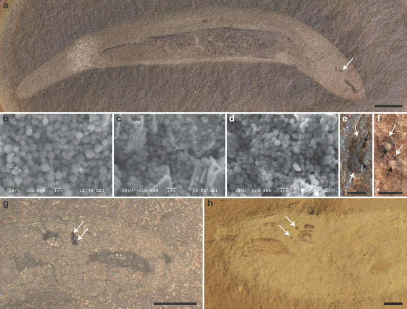
The hagfish eye is highly reduced, lacking pigment and a lens, and covered by soft tissue. The timing and mode of this loss of complexity remain unknown. Here, we present high-resolution anatomical data on the fossilized eyes of three stem hagfishes that form a transitional series towards the highly vestigialized eye of modern hagfishes. All have small eyes covered in soft tissue and containing only spherical melanosomes. Lenses are present in the more stemward hagfishes but absent in the more crownward hagfish. Reduction of the eyes occurred gradually across the Palaeozoic, with an initial stage of size reduction and loss of cylindrical melanosomes from the retinal pigmented epithelium, an intermediate stage with loss of image focus capability and finally a near-complete loss of vision (the hagfish crown group). The initial and intermediate stages of this process likely occurred in nearshore environments prior to the Permian colonization of the continental slope by hagfish.


The Lauer Foundation was involved in a study of hagfish eyes with collaborators from the University of Wisconsin- Milwaukee, The Sandra and David Douglass Collection, Field Museum and the University of Ottawa published in the journal Biology Letters.
Figure 1. Fossil specimens of Gilpichthys greenei housed in the collections of either the Lauer Foundation for Paleontology, Science and Education (specimen numbers starting with LF) or the Field Museum of Natural History (specimen numbers starting with FMNH). (a) Specimen LF 6966 showing the entire body with the eyes (essentially overlapping) indicated by a white arrow; scale bar = 5 mm. (b–d) SEM secondary electron images of the eyes of three specimens of G. greenei, showing preserved melanosomes. (b) Specimen LF 6965. (c) Specimen LF 6970. (d) Specimen LF 6967. (e) Specimen LF 6968 showing eyes (white arrows) with lenses preserved as dark holes (black arrows); scale bar = 0.5 mm. (f) Specimen LF 6966 eyes (white arrows) with lenses preserved in white kaolinite (black arrows); scale bar = 0.5 mm. (g) Specimen LF 6965 preserved in almost direct lateral view, with eyes overlapping, and cranial elements posterior to their eyes preserved as a line; scale bar = 2 mm. (h) Specimen FMNH PF8348 preserved offset from lateral, with widely spaced eyes and cranial elements preserved posterior to the eyes as wideset structures; scale bar=2mm

Press Coverage:


Comments


Commenting on this post isn't available anymore. Contact the site owner for more info.
bottom of page